Radionuclide Imaging and Therapy
What are radionuclides?
Radionuclide Imaging and Therapy: What You Need to Know
Radionuclides just radioactive versions of regular elements. Some are made by humans (like during nuclear power generation) and some are found in nature. But what's really cool is that each one gives off radiation at its own speed - this is measured by something called half-life. And since each radionuclide is different, they all give off radiation at different rates.
Radionuclide Imaging and Therapy: Half-life
Understanding Half-Life and Radioactive Decay
In radioactivity half-life is a really important concept. It's the time it takes for half of the atomic nuclei in a radioactive sample to decay. When this happens, particles and energy are released, and the radioactive substance transforms into something else. This is known as decay. The average time it takes for the number of unstable nuclei to halve is called the half-life of an isotope.
Radionuclide imaging Techniques
There are three techniques of radionuclide imaging used in today's medical physics. They are planar scintigraphy, also known as single-photon emission computed tomography (SPECT), positron emission tomography (PET), and hybrid techniques.
Planar scintigraphy or single-photon emission computed tomography
Gamma-emitting radionuclides are commonly used in medical imaging for the detection of various diseases and disorders. Planar imaging and single-photon emission computed tomography (SPECT) are two widely used techniques that require radiopharmaceuticals containing gamma-emitting radionuclides.
Both methods rely on gamma cameras for detection, which use collimated detectors to register emitted gamma rays. A series of collimators direct gamma rays into an array of scintillation crystals, which transform them into optical photons and detect them with photomultiplier tubes (PMT). Using this data, a two-dimensional image (scintigram) of radioactivity distribution is created.
SPECT has the advantage of three-dimensional imaging (tomography) for better data. Coll describe the optimum energy as being between 140 and 160 keV. The most frequent gamma-emitting radionuclides utilized in planar scintigraphy and SPECT are 99mTc, 111In, and 123I. The numbers 99m, 111, and 123 indicate the mass number, also known as the nucleon number, and can be calculated by the addition of protons and neutrons.
To minimize unwanted irradiation, the radionuclide's half-life should be short enough to fade away as soon as possible after imaging. Overall, the use of gamma-emitting radionuclides in medical imaging has greatly improved the diagnosis and treatment of various diseases and disorders.
Positron emission tomography
Positron emission tomography (PET) is a unique and non-invasive technique used to monitor and quantify molecular interactions in vivo. PET detects the regional concentration of an imaging agent, providing real-time data on physiological processes.
PET requires the use of a radiopharmaceutical containing a positron-emitting radionuclide. These radionuclides require an extra neutron to attain a lower energy state. To achieve stability, they undergo spontaneous decay, producing a neutron and emitting a positron and a neutrino.
When the radiopharmaceutical is administered to the patient, the positron emitted by the radionuclide interacts with an electron in the body, resulting in the production of two gamma rays. These gamma rays are detected by the PET scanner, which produces a three-dimensional image of the distribution of the radiopharmaceutical in the body.
PET is a highly precise technique, allowing for the detection and quantification of molecular interactions in real-time. It is commonly used in the diagnosis and treatment of various diseases, including cancer, Alzheimer's disease, and heart disease. Overall, PET hasized medical imaging by accurate-in method for physiological processes in vivo.

The positron travels a given distance (positron range), which is determined by the density of the environment and the positron energy. When its kinetic energy drops, it makes contact with an electron, resulting in its annihilation and the creation of two 511 keV photons. After that, PMT registers the photon counts. Single photon events are rejected, allowing for precise measurement of the radioactivity content in the target region. The registered events are rebuilt into pictures that represent the radioactive source's spatial distribution throughout the body.
It is possible to do recurrent examinations during the same day because of an isotope with a short half-life of up to 68 minutes (68Ga).
Radionuclide Imaging and Therapy: Hybrid techniques
The addition and integration of computed tomography (CT) to SPECT and PET for the acquisition of morphologic information while the patient is in the same position resulted in further development and improvement of the nuclear imaging technique. This is critical for pinpointing the exact location of the lesions, particularly in the abdominal region.
Imaging processes are shortened with the use of a CT attenuation map. PET-CT is a hybrid imaging technique that combines the sensitivity of PET with the temporal and spatial resolution of CT. PET and SPECT quantification accuracy is also improved by CT attenuation and scatter correction.
A PET-CT scan. flickr.com
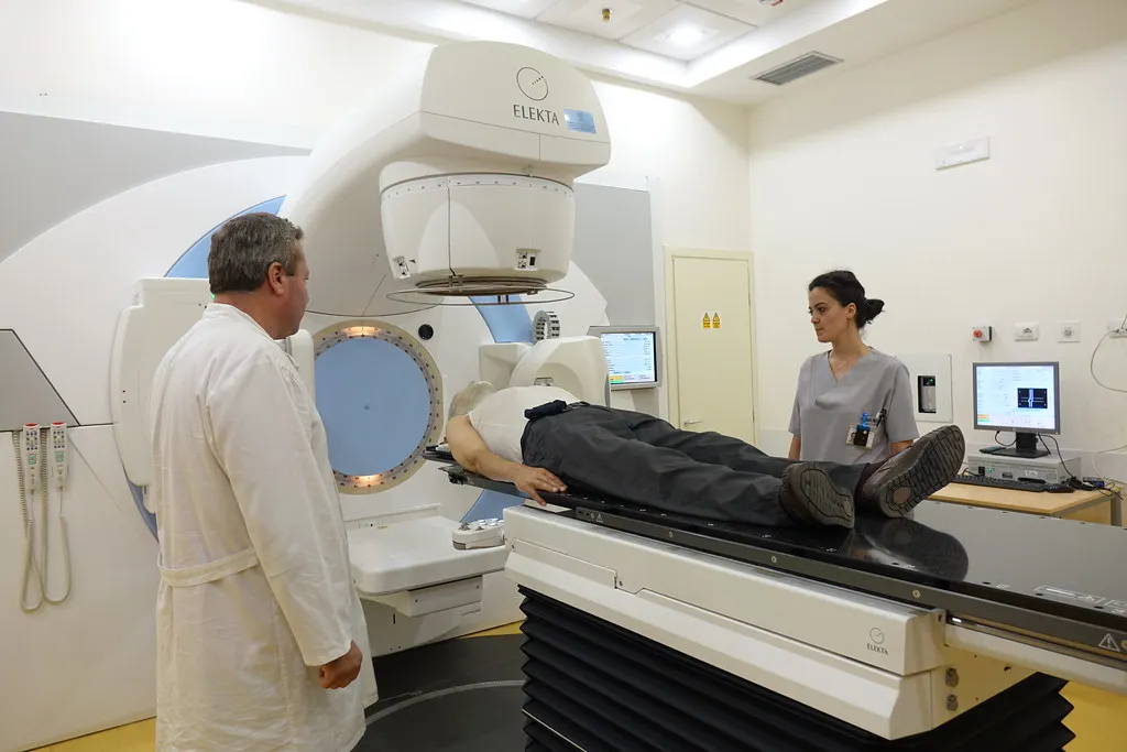
Radiotherapy
Ionizing radiation is a powerful tool in the fight against cancer. It is highly effective in regulating or destroying fast-dividing cancer cells due to their sensitivity to radiation. External and internal radiotherapy are both possible, with the latter involving the use of sealed and implanted radiation sources (brachytherapy) or intravenously supplied radionuclides for in vivo molecular interaction during internal radiotherapy.
The role of radiotherapy in nuclear medicine is becoming increasingly important. Targeted radiotherapy of small tumors, micrometastases, and single cancer cells can all benefit from radionuclides Auger theular range. These radionuclides differ in terms of the type of radiation they emit, as well as their radiobiological efficacy and range of action, allowing for tumor type selection.
Despite its effectiveness, radiation therapy can have adverse effects on healthy cells, leading to tissue damage and other side effects. Therefore, it is crucial to carefully plan and monitor radiation therapy to minimize these risks and optimize the therapeutic benefits for each patient. With advances in technology and research, radiotherapy is becoming an increasingly important component of cancer treatment, offering new hope for patients and their families.
Radiotherapy has either found an application or shown potential in solving lymphoma, breast, prostate, colon, thyroid, lung, and brain cancer types, as well as in bone pain palliation.
Radionuclide Imaging and Therapy - Key takeaways
Understanding the half-life of a radioactive sample is crucial in the field of nuclear medicine. It refers to the time it takes for one-half of the atomic nuclei to decay, which explains how nuclear species can transition spontaneously into other nuclear species by releasing particles and energy. The radioactive versions of elements are called radionuclides, which can be found in nature or created by humans through nuclear processes.
In medical physics, radionuclide imaging plays a vital role in diagnosing, staging, and monitoring diseases such as cancer, trauma, and infection. Three techniques used in radionuclide imaging are single-photon emission computed tomography (SPECT), positron emission tomography (PET), and hybrid techniques. The choice of technique depends on the specific medical condition and the imaging requirements.
The half-life of a radionuclide used in imaging must be short enough to reduce unwanted irradiation and fade away as soon as possible after imaging. This reduces the risk of harmful radiation exposure and allows for more frequent imaging if needed.
Overall, radionuclide imaging is a powerful tool in medical physics that allows for non-invasive and highly accurate diagnosis and monitoring of diseases. As technology and research continue to advance, radionuclide imaging is likely to become even more effective in improving patient outcomes and quality of life.
Radionuclide Imaging and Therapy
Why is the value of a radionuclide's half-life important?
The value of a radionuclide's half-life is important because it determines if it can be used in radionuclide imaging. If it is short enough, it allows for repeated examinations during the same day.
What is radiotherapy?
The use of strong doses of radiation to destroy cancer cells and reduce tumors is known as radiotherapy.
What is radionuclide therapy used for?
Targeted radiotherapy of small tumors, micrometastases, and single cancer cells can all benefit from radionuclides that produce Auger electrons in the subcellular range. Radionuclides differ in terms of the type of radiation they emit, as well as their radiobiological efficacy and range of action, allowing for tumor type selection. Also, it has either found an application or shown potential in solving lymphoma, breast, prostate, colon, thyroid, lung, and brain cancer types, as well as in bone pain palliation.


